Thursday, July 20, 2006

HIV Genome


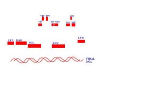
The HIV genome is made up of three genes that code for inner core proteins, envelope proteins and functional proteins (i.e. viral proteins). These are named gag (group antigen-since the antigenicity of this inner core is conserved through out the group, env (envelope) and pol (polymerase) gene which codes for Reverse Transcriptase which has both polymerase and ribonuclease activity. It also codes for other enzymes like integrase whose function is to facilitate the integration of viral DNA into the host cell chromosome and protease which cleaves precursor proteins into smaller functional fragments-the envelope glycoprotein is made as gp160 but is cleaved to form gp120 and gp41.


As with other retroviruses, HIV has at the end of its genome a segment of nucleic acid called LTR (Long Terminal Repeats) which are stretches of DNA that do not by itself code for any protein but functions as a regulator to the expression of the three structural genes, a function that is augmented by other genes called regulatory genes.


Regulatory Genes

These regulate the production of viral proteins: one regulator speeds up protein synthesis, another speeds the production of only some kinds of proteins and a third represses protein synthesis. Each regulatory gene encodes a protein that interacts specifically with a responsive element which is a sequence of nucleotides within the genome. Regulatory genes act in trans because they exert at a distance, the responsive sequence affect adjacent genes hence act in cis. Through their influence regulatory genes can effect an explosive viral replication, moderate growth or quiescence.

The tat Gene

That tat gene (Trans-activator of transcription) is both unusual in both structure and effect; it occurs as two widely separated sequences of nucleotides. The gene starts from nucleotide 5831 to 6045 and then 8379 to 8424.

This gene is translated to a polypeptide called Tat. This protein when bound to the viral RNA boost the expression of viral genes. It interacts with a kinase that is involved in the phosphorylation of the carboxy-terminal which encode a protein which when bound to viral RNA boost the expression of viral gene domain (CTD) of RNA polymerase-II required for initial transcription. To exert its effect the tat protein is dependent upon a short sequence of nucleotides known as TAR (trans-activating responsive sequence). Tat stimulatory effect extend to all viral proteins, it also positively feeds back upon itself resulting in enormous synthesis of viral proteins. It activates the expression of TNF-β, TGF-β but down-regulate other cellular gene expression including bcl-2 and the chemokine MIP-1α.


The rev Gene

A second regulator gene rev (regulator of expression of viral-protein) also exists as two widely separated nucleotide sequence, one from 5 970 to 6 045 then another from 8 379 to 8 653. The gene encodes a 13kD protein Rev which enables the integrated virus to produce selectively either regulatory proteins or virion components. It acts as a genetic switch whose role is in the activation of the virus from latency to active viral replication.

The vpr Gene

The gene is found between the 5 559th and 5 795th nucleotide. It encodes a Vpr protein. The protein is incorporated into viral particle. Approximately 100 copies of vpr are associated with each virion. This is mediated through specific interaction between the carboxyl terminal region of p55 gag which corresponds to p6 in the proteolytically processed protein. It confers the ability of HIV-1 to infect non-dividing cells by facilitating nuclear localisation of the pre-integration complex. This NL is distinct from the prototypic NLS because there is no negative feedback, but the NLS peptide can inhibit the nuclear localisation of other proteins containing conventional NLS eg matrix (p17) protein. It also affect cell division- all cells expressing vpr accumulate in the G2 phase of the cell cycle; vpr expression prevents activity of p34cdc2/cyclin B complex an activator of the cell cycle important for entry into mitosis.

The vpu gene (6062-6310)

Vpu codes for a 16kD polypeptide- an integral membrane phosphoprotein localised in the internal membrane of the cell. It is expressed from mRNa that also encodes for envelope proteins but translated at levels ten times lower than that of env. A complex gp120/CD4 occurs in the endoplasmic reticulum but vpu degrades the CD4 molecule because this complex interferes with virion assembly.

The vif Gene (5 041- 5 619)

Codes for a 23kD protein which is responsible for the replication of HIV in the peripheral blood lymphocytes & macrophages. Other cells have complimentary proteins to vif hence not needed.

The nef Gene (8 797-9168)

The Nef gene (negative –regulator factor) encodes a 27kD myristylated protein which is the first viral protein to accumulate to detectable levels in cells following HIV-1 infection. The protein down-regulates the expression of CD4 by infected cells by stimulating CD4 endocytosis and lysosomal degradation by interaction with the dileucine repeat sequence contained in its membrane proximal region. Also down-regulates cell-surface expression of MHC-1, this is the protein that is responsible for the perturbation of T-cell activation and stimulation of infectivity.


© freeman Chari 2005

Monday, July 17, 2006

Origins Of HIV

So many questions have been posted since the discovery of the Human Immunodeficiency Virus but the most compelling has been that of its origins. Is it a new virus or an old virus that had lied dormant but going through mutations?

One way of tracing its origins was to look for similar viruses in non-human primates. Monkeys and apes were the most notable targets since they are known to be infected by some other important viruses that are known to infect man e.g. yellow fever virus.

In 1984 Max Essex et al examined large numbers of primates by serologically testing for cross-reactive epitopes in their sera. They found thus a monkey virus related to HIV in blood samples of the Asian macaques (macaca mulatta) housed at New England primate centre. At the same time captive macaques were reported to be developing AIDS-like symptoms, the virus was isolated and named SIV (simian immunodeficiency virus). It was clearly related to HIV; it infected the same CD4 subset of lymphocytes and reacted with antibodies produced by AIDS patients. The biochemical properties of SIV were similar to those of HIV.

Further studies have shown that SIV and HIV are 50% related at nucleotide level and that the organisation of structural and regulatory genes except that SIV has vpx gene which is not found in HIV and equally HIV has vpu gene not found in SIV. Like in humans with HIV, SIV infected monkeys showed decreased T4-cell count with evident immunosuppression and subsequent death to opportunistic infection.

What shocked researchers was that wild Asian macaques did not have any evidence of prior exposure to SIV or any HIV-like agent, thus infection had been limited to a small group of captive monkeys. This suggested that SIV did not naturally infect Asian monkeys in the wild.

In the same year blood samples from representative African primates, including wild caught chimpanzees (pan troglodytes); African green monkeys (cercopithecus aethiopis) and baboons (papio spp) were analysed. No evidence of SIV was found in chimpanzees or baboons but 50% of the African green monkeys did show evidence of SIV infection. Subsequent analysis of samples from other African Green monkeys has shown that 30-70% have SIV infection.

What puzzled investigators was why SIV does not seem to cause any disease or harm to AG monkeys yet wrecked havoc to Asian monkeys. This observation has also been made in that chimpanzees are the only animals that can be experimentally infected with HIV isolated from AIDS patients, yet the virus does not seem to cause any lethal disease in chimpanzees as it does in man.

Clearly SIV is the closest relative of the Human Immunodeficiency Virus, however the 50% relationship at nucleotide sequence level could not make it the immediate precursor of HIV, thus probably there were other intermediate viruses. In 1985 there was evidence in Senegal of such a virus.

It was found that about 10% of prostitutes there had antibodies that reacted with both HIV and SIV. Surprisingly the antibodies reacted much better with SIV than HIV particularly the external envelop glycoproteins. HIV-positive sera from US and central Africa did not react very well with the SIV envelope antigens. This virus was then called HIV-2, nevertheless studies have shown that people with HIV-2 have antibodies that are entirely reactive with SIV antigens. Genetic studies have shown that their nucleotide sequences are closely related too suggesting that these viruses share evolutionary roots.

Essex’s study showed that HIV-2 was endemic in West Africa. Isolation of HIV-2 from patients referred to Europe for treatment suggested that HIV-2 could in fact cause AIDS. Seroepidemiological studies showed that HIV-2 was transmitted sexually. Prostitutes who tested HIV-2 positive in 1985 were subsequently followed and examined for any abnormal clinical symptoms. In contrast to prostitutes in other parts of Africa with HIV-1, they showed negligible rates of lymphadenopathy, AIDS-related complex or AIDS itself. Thus the virulence of HIV-1 was greater as compared to HIV-2.

Friday, July 07, 2006

HIV & AIDS- THE HISTORY

Between October 1980 and May 1981 Dr Michael Gottleib and his colleagues in Los Angeles were perplexed by a cluster of male patients whose age ranged from 29 to 36 years under their care and surveillance. All five men were diagnosed as infected by pneumocystis carinii and all had pneumocystis carinii pneumonia (PCP) a disease previously exclusively associated with severely immunosuppressed patients, in addition all of them had evidence of exposure to cytomegalovirus (CMV) a virus also commonly seen in immunosuppression. Furthermore, all had thrush, a candidal infection only present in frail immunity. In three of these five cases there was evidence of marked disturbance to the functional capabilities of their immune system. Another notable feature was on the five was that all were sexually active homosexuals. None of them knew each other and there was no common sexual contact between them.

In July 1981 a similar report was made of 26 homosexual men from New York and California. They, in addition had an uncommon tumour called Kaposi’s Sarcoma which had been previously observed in elderly men of Jewish origin and in Tropical Africa mainly in children and young adults.

These cases although few made an impact because the disease was obviously transmissible from person to person and because of its evident effect on the immune system of patients it became known as Acquired Immunodeficiency Syndrome (AIDS).

In America this was a disease associated with homosexuals and intravenous drug users but African patients with AIDS appeared to lack these two lifestyle risk factors; it also differed with respect to the sexuality of the infected. In fact it was found to be equally distributed in both males and females.

The disease was then clearly described with a set of clinical criteria established which could be used to define the infection, but what could not be ascertained was the primary cause of the disease. With the characteristic association with homosexuality it was therefore logical to search for clues amongst people with this kind of lifestyle. One early theory was the use of amyl nitrite a chemical used commonly as a reliever of angina due to coronary artery constriction since it is a vasodilator. Homosexuals used it to increase penile erection; however, it was known to be a relatively strong immunosuppressor, hence the belief that continuous use resulted in gradual progressive destruction of the immune system.

Another possibility was that of cytomegalovirus (CMV); a virus that infects humans and so called because it caused infected cells to become pathologically enlarged. The basis of this theory was that virtually all patients with AIDS had evidence of cytomegalovirus and may suffer from the diseases characteristic of its infection like pneumonia, retinal infection and diarrhoea. In addition one of its important routes of transmission is venereal. It was however discovered that not all patients with CMV infection had signs of immunosuppression and CMV, as characteristic of all members of the Herpes Virus family (herpersviridae) could exist in a latent form and only produced symptoms when activated. Immunosuppression is one of the triggering factors.

The link between Kaposi’s sarcoma and AIDS brought another dimension on the ever increasing number of possibilities. For long retroviruses had been known to have cancer-causing potential. It had also been shown that some retroviruses isolated in animals caused leukaemias as well as solid tissue tumors, hence KS could also be another disease caused by retroviruses.

The discovery of reverse transciptase- an enzyme responsible for the formation of a DNA copy from an RNA template- in the mid 1970s by H M Temin and (independently) David Baltimore brought to light the replication and life cycle of retroviruses. At the about the same time Gallo and colleagues at the National Institute of Health (USA) discovered a growth factor for mature lymphocytes which would permit them to grow in vitro. This was an important development because it made it possible to grow lymphotropic viruses in culture. However despite such importantdiscoveries no infectious retroviruses had been isolated and investigators believed that no human retroviruses existed. This scepticism was largely due to the fact hat it had been largely easy to isolate animal retroviruses so was the expectation for human retroviruses. Gallo in 1980 managed to isolate the first human retrovirus which he named: Human T-lymphotropic Virus Type one ( HTLV-1)

HTLV-1 to date still remains the most notable tumor virus definitively established to be a causal of human malignancy. It infects the white blood cells and causes a rare but highly malignant cancer of the leukocytes called Adult T-cell Leukemia (ATL) mostly common in Japan and Africa.

Two years later they isolated another closely related virus which they named HTLV-II nd it is thought to cause hairy-cell leukaemia. These two viruses however showed four crucial features: that they are spread by blood, sexual contact, mother-to-child and that they both infected T-lymphocytes.

The discovery of AIDS projected an additional significance of the two retroviruses. There were two important observations : AIDS was defined as immunosuppression and that it could be transimitted by sexual and blood contact. HTLV-1 had been shown to use the same route, furthermore Max Essex of Harvard School of Public Health had shown that a retrovirus confined to cats called feline leukaemia virus (FeLV) could cause both cancer and immunosuppression. Thus, the hypothesis held then that the cause of AIDS was a close relative of HTLV-1.

In 1983 Dr Francoise Barre-Sinoussi together with Luc Montagnier of the Pasteur Institute analysed tissues from a patient with lymphadenopathy syndrome. The specimen was minced and put in tissue culture whence they tested for reverse transcriptase. The activity was found to be there but further tests showed that the retrovirus was neither HTLV-1 nor HTLV-II. They named it Lymphadenopathy-associated Virus (LAV). It grew in T4-cells ( T cells with a CD4 cell marker) but not in T8 cells. It was shown that the virus could kill or inhibit T4 cell growth.

Robert Gallo, the following year propagated the virus in cell culture and called it HTLV-III. The virus was hitherto known as HTLV III/ LAV to give equal recognition to both camps. To compound the already existing confusiona virus isolated from an AIDS patient by Dr Jay Levy was given yet another name, ARV (AIDS-related virus). It was however shown after gene probing that these viruses are identical although with a tolerable difference. Thus an international committee after formal agreement between President Reagan of USA and President Mitterand of France decided to change the name to Human Immunodeficiency Virus (HIV).

In 1986 Luc Montagnier’s group isolated another strain which they called LAV-2 from patients in Guinea Bissau and the Cape Verde. At about the same time an American group under Essex isolated a second virus in Senegal which they called HTLV-IV, but these were again shown to be identical and were later called HIV-2.